octubre 2025 - DERMAGIC EXPRESS / Dermatologia y Bibliografia - Dermatology & bibliography DERMAGIC EXPRESS / Dermatologia y Bibliografia - Dermatology & bibliography: octubre 2025

viernes, 24 de octubre de 2025

THE SEBORRHEIC DERMATITIS, A REVIEW. / LA DERMATITIS SEBORREICA, REVISION


LA DERMATITIS SEBORREICA Y SUS ALTERNATIVAS TERAPÉUTICAS, ACTUALIZACIÓN.


THE SEBORRHEIC DERMATITIS AND THERAPEUTIC ALTERNATIVES, A REVIEW.


DERMATITIS SEBORREICA MEJILLAS


Dermatitis seborreica

PUBLICADO 2017 ACTUALIZADO 2025


EDITORIAL ESPAÑOL
===================
Hola amigos de la red, DERMAGIC EXPRESS de nuevo, hoy con el interesante tema: LA DERMATITIS SEBORREICA, una enfermedad no tan severa pero muy frecuente hoy en dia en la población, tanto niños como adultos, hombres y mujeres.
 
 
1.) HISTORIA:
 
La dermatitis seborreica fue descrita por primera vez en 1732 por Paul Gerson Unna,  medico Alemán, uno de los pioneros en dermatología. 
 
En 1873 el medico Italiano Giuseppe Rivalta sugirió un posible origen micótico de la DERMATITIS SEBORREICA.
 
En 1874 Louis-Charles Malassez medico oriundo de Francia, relacionó la presencia de levaduras llamadas Malassezia con la dermatitis seborreica, al identificar unas estructuras fungicas en el cuero cabelludo las cuales denomino "champignon de la pelade" ("hongo de la calvicie"), y las relaciono con la pitiriasis capitis (una afección ligada a la dermatitis seborreica). 
 
Su contribución fue importante y diriase clave al descubrir la presencia de estas levaduras en la piel, y su probable papel en algunas enfermedades dermatológicas. debido a este hecho, el género de estas levaduras fue nombrado "Malassezia" en su honor,
 
En 1952 Riccardo Galeazzi Leone medico italiano tambien,  encontró la asociación entre Pityrosporum ovale (hoy Malassezia furfur) y esta enfermedad.
 
Con respecto al hongo MALASSEZIA FURFUR, esta fue descubierta en 1846 por el médico ginecólogo y dermatólogo alemán Karl Ferdinand Eichstedt, relacionada con LA PITIRIASIS VERSICOLOR, también patología cutánea; y posteriormente también se le relaciono con la DERMATITIS SEBORREICA, como lo acabamos de mencionar.

2.) SÍNTOMAS CLÍNICOS:

La enfermedad afecta principalmente la cara, el cuero cabelludo, y también tórax anterior y posterior menos frecuentemente. 
 
Caracterizada por ser placas eritematosas y descamativas con prurito, es de evolución crónica y representa un verdadero reto su tratamiento, debido al hecho de que es frecuente su recaída, aun con las mejores opciones de tratamiento

Las areas mas afectadas del organismo son las llamadas "zonas seborréicas" que son las áreas del cuerpo con mayor concentración de glándulas sebáceas y donde suele aparecer la dermatitis seborréica, incluyen:

A - Cuero cabelludo: la línea de implantación del cabello, llamada corona seborréica, pero todo el cuero cabelludo puede estar afectado.

B - Región facial media:  cejas, surcos nasogenianos, alas de la nariz.
 
C - Región del pecho medio y zona interescapular de la espalda: entre  omóplatos.
 
D - Detrás de las orejas y conductos auditivos externos.
 
E - Pliegues axilares, submamarios e inguinales: no tan frecuente.

F   - Área periglútea: pliegue interg
teo.

G - Párpados y borde palpebral:  a veces afectando las pestañas, conocida con el nombre de BLEFARITIS SEBORREICA.

Estas zonas se caracterizan por presentar piel grasa, o con secreción sebácea abundante, lo cual favorece el desarrollo de la enfermedad.

En el cuero cabelludo la descamación producida por la DERMATITIS SEBORREICA es conocida con el nombre vulgar de "CASPA", quizá una de las causas mas frecuentes de la consulta dermatologica.


DERMATITIS SEBORREICA DE LA CARA


Dermatitis seborreica de la cara


3.) EVOLUCIÓN:

El Curso de la DERMATITIS SEBORREICA es crónico y recurrente, con períodos de remisión y exacerbación. 
 
Puede comenzar desde la infancia, y el rango mas comun esta en adultos jóvenes y mayores. 
 
Los brotes empeoran con estrés, clima frío o muy calurosoy enfermedades subyacentes (p. ej. VIH, Parkinson). En raros casos, en algunos pacientes puede generalizarse, pero suele limitarse a zonas seborreicas ya descritas.

4.)  TRATAMIENTOS:
 
A.- Decada de los años 50-60: comenzaron a utilizarce los corticosteroides topicos para disminuir la inflamacion Uso de corticosteroides tópicos, de baja potencia (HIDROCORTISONA) , la SULFACETAMIDA, y el AZUFRE, para disminuir la el prurito, eritema e inflamación, aun vigentes hoy dia.
 
B.- En la decada de los años 70-80: se introdujeron FORMALMENTE el uso de antimicoticos como KETOCONAZOL, FLUCONAZOL, BIFONAZOL, ITRACONAZOL, SERTACONAZOL, MICONAZOL, TREBINAFINA y CICLOPIROXOLAMINA, para controlar la levadura MALASSEZIA, algo tardio, pues la relacion de este hongo con la dermatitis seborreica se establecio practicamente en los años 50.
 
- En los eños 80 (1985), se introdujo ESPECIFICAMENTE el ITRACONAZOLE.
- En los alos 90 se comenzo a usar el METRONIDAZOLE en gel topico. 
 
C.- Decada de los años 90 - 2000: se introduce  el uso adicionalde queratoliticos y alquitranes (aceite de cade, urea) para mejorar la descamacion y producir la regeneracion cutanea.
 
- En los años 2000 aparecen el TACROLIMUS y PIMECROLIMUS, como alternativas terapeuticas. 
 
D.- Decadas de los años 2000 -2025: Se introduce la combinacion de los anteriores: antimicoticos topicos, con cosrtoesteroides de baja potencia, y formulas magistrales con bioasufre fluido, ciclopiroxolamina, y otros componentes,  preparadas en champu y cremas para uso topico.


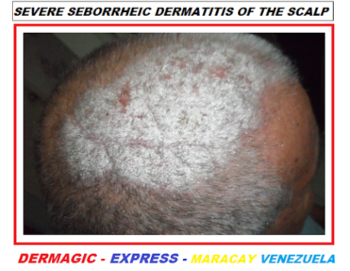
DERMATITIS SEBORREICA SEVERA DEL CUERO CABELLUDO


Dermatitis seborreica cuero cabelludo


Hoy dia 2.025 les presento en esta revisión las MEJORES ALTERNATIVAS TERAPÉUTICAS que han sido descritas en el tratamiento de esta enfermedad: 

1.) CORTICOSTEROIDES TOPICOS:  CLOBETASOL,  DEXAMETASONA, MOMETASONA.
2.) UREA.
3.) ÁCIDO SALICÍLICO.
4.) PROPILENGLICOL.
5.) PIRITIONATO DE ZINC.
6.) BIOTINA.
7.) KETOCONAZOL.
8.) FLUCONAZOL.
9.) SERTACONAZOL.
10.) ITRACONAZOL.
11.) DITRANOL.
12.) SUCCINADO DE LITIO.
13.) VITAMINA-COMPLEJO B.
14.) PEROXIDO DE BENZOILO.
15.) SULFURO DE SELENIO.
16.) SULFACETAMIDA.
17.) TACROLIMUS y 18.) PIMECROLIMUS  (Hoy día 2025 estos medicamentos tienen una advertencia severa por la FDA tipo "BLACK BOX" o "CAJA NEGRA", en sus enpaques acerca de que el prolongado puede inducir malignidad)
18.) PIMECROLIMUS.
19.) METRONIDAZOL.
20.) TERBINAFINA.
21.) ALQUITRAN DE PINO.
22.) ALQUITRAN DE HULLA.
23.) GLICERINA.
24.) TACALCITOL.
25.) FOTOTERAPIA y FOTOQUIMIOTERAPIA
26.) AZUFRE.
27.) BIFONAZOL.
28.) CICLOPIROXOLAMINA.
29.) L - DOPA.
 
30.) SULFATO o GLUCONATO DE ZINC.

Probablemente existan  otras mas, de modo que el tratamiento de la DERMATITIS SEBORREICA hoy dia, constituye un RETO para todo dermatólogo, pues el indice de RECAÍDAS o BROTES es muy FRECUENTE.

5.) NUEVOS TRATAMIENTOS:

- ROFLUMILAST: es un inhibidor de la fosfodiesterasa-4 (PDE4) cuyo mecanismo de accion es reducir la inflamación cutánea. 
 
- Se trata de una espuma tópica  al 0,3% la cual fue aprobada el 16 de diciembre de 2023 por la FDA para el tratamiento de la DERMATITIS SEBORREICA, en adultos y niños mayores de 9 años de edad.
 
- Es el primer medicamento aprobado en más de dos décadas, quiere decir esto desde los años 80, para tratar esta patología; su principal efecto: controlar el enrojecimiento y la y la inflamación cutaea.

De resto se siguen utilizando las ya conocidas, cremas y champu comerciales, quedando como GRANDES opciones LAS FORMULACIONES MAGISTRALES para estos casos, como champús preparados a base de azufre, ácido salicílico y lociones capilares también preparadas, INTERCALADAS con productos ya conocidos.

La dermatitis seborreica en muchos casos es recalcitrante, difícil de tratar pues recaen mucho los pacientes una vez presentan mejoría, el exceso de grasa en la cara y el estrés son GRANDES factores desencadenantes y de recaídas.

Saludos a todos. 

Dr. José Lapenta.
Dr.  José M. Lapenta.


EDITORIAL ENGLISH
===================
Hello friends of the network, DERMAGIC EXPRESS is back again, today with the interesting topic: SEBORRHEIC DERMATITIS, a disease that is not very severe but very common nowadays in the population, affecting both children and adults, men and women.

1.) HISTORY:


SEBORRHEIC DERMATITIS was first described in 1732 by Paul Gerson Unna, a German physician and one of the pioneers in dermatology.

In 1873, the Italian physician Giuseppe Rivalta suggested a possible fungal origin for SEBORRHEIC DERMATITIS.

In 1874, Louis-Charles Malassez, a physician from France, linked the presence of yeasts called Malassezia to seborrheic dermatitis. He identified fungal structures on the scalp, which he called "champignon de la pelade" ("baldness fungus"), and associated them with pityriasis capitis (a condition linked to seborrheic dermatitis).

His contribution was significant and arguably key in discovering the presence of these yeasts on the skin and their probable role in some dermatological diseases. Because of this, the genus of these yeasts was named "Malassezia" in his honor.

In 1952, Riccardo Galeazzi Leone, also an Italian physician, discovered the association between PITYROSPORUM OVALE (now MALASSEZIA FURFUR) and this disease.

Regarding the fungus MALASSEZIA FURFUR, it was discovered in 1846 by the German gynecologist and dermatologist Karl Ferdinand Eichstedt, and was associated with PITYRIASIS VERSICOLOR, also a skin condition; and later it was also linked to SEBORRHEIC DERMATITIS, as we just mentioned.

2.) CLINICAL SYMPTOMS:

The disease primarily affects the face and scalp, and less frequently the anterior and posterior chest.

Characterized by itchy, scaly, erythematous plaques, seborrheic dermatitis is a chronic condition that presents a significant treatment challenge due to its frequent relapse, even with the best treatment options.

The most affected areas of the body are the so-called "seborrheic zones," which are areas with the highest concentration of sebaceous glands and where seborrheic dermatitis typically appears. These include:

A - Scalp: the hairline, called the seborrheic corona, but the entire scalp can be affected.

B - Midface: eyebrows, nasolabial folds, and sides of the nose.

C - Mid-chest and interscapular region of the back: between the shoulder blades.

D - Behind the ears and external auditory canals.

E - Axillary, inframammary, and inguinal folds: less common.

F - Perigluteal area: intergluteal fold.

G - Eyelids and eyelid margins: sometimes affecting the eyelashes, known as SEBORRHEIC BLEPHARITIS.

These areas are characterized by oily skin, or abundant sebum secretion, which favors the development of the disease.

On the scalp, the flaking caused by SEBORRHEIC DERMATITIS is commonly known as "DANDRUFF", perhaps one of the most frequent reasons for dermatological consultations.
 
3.) EVOLUTION:

The course of seborrheic dermatitis is chronic and recurrent, with periods of remission and exacerbation.

It can begin in childhood, and it is most common in young adults and older adults.

Outbreaks worsen with stress, cold or very hot weather, and underlying diseases (e.g., HIV, Parkinson's). In rare cases, it can become generalized in some patients, but it is usually limited to the seborrheic areas already described.

4.) TREATMENTS:

A.- 1950s-1960s: Topical corticosteroids began to be used to reduce inflammation. The use of low-potency topical corticosteroids (hydrocortisone), sulfacetamide, and sulfur to reduce itching, erythema, and inflammation is still current.

B. In the 1970s and 80s: the use of antifungals such as KETOCONAZOLE, FLUCONAZOLE, BIFONAZOLE, ITRACONAZOLE, SERTACONAZOLE, MICONAZOLE, TERBINAFINE and CYCLOPRIROXOLAMINE was FORMALLY introduced to control the MALASSEZIA yeast. This was somewhat late, as the relationship between this fungus and seborrheic dermatitis was practically established in the 1950s.

- In the 1980s (1985), ITRACONAZOLE was specifically introduced.

- In the 1990s, METRONIDAZOLE began to be used in topical gel form.

C.- From the 1990s to 2000: the additional use of keratolytic agents and tars (cade oil, urea) was introduced to improve scaling and promote skin regeneration.

- In the 2000s, TACROLIMUS  and PIMECROLIMUS emerged as therapeutic alternatives.

D.- 2000-2025: Combinations of the above were introduced: topical antifungals with low-potency corticosteroids, and MAGISTRAL FORMULATIONS with fluid biosulfur, ciclopiroxolamine, and other components, prepared as shampoos and creams for topical use. 


soborrheic dermatitis of the scalp
 
 
Today, in 2025, I present in this review the BEST THERAPEUTIC ALTERNATIVES that have been described in the treatment of this disease:
 
1.) TOPICAL CORTICOSTEROIDS: CLOBETASOL, DEXAMETASONE, MOMETASONE.
2.) UREA.
3.) SALIYLIC ACID.
4.) PROPYLENE-GLYCOL.
5.) PYRITHIONE ZINC.
6.) BIOTIN.
7.) KETOCONAZOLE.
8.) FLUCONAZOLE.
9.) SERTACONAZOLE.
10.) ITRACONAZOLE.
11.) DITHRANOL.
12.) LITHIUM SUCCINATE.
13.) VITAMIN-COMPLEX B.
14.) BENZOYL PEROXIDE.
15.) SELENIUM SULFIDE.
16.) SULFACETAMIDE.
17.) TACROLIMUS. 18.) PIMECROLIMUS, (Today 2023 these drugs TACROLIMUS AND PIMECROLIMUS have a severe warning from the FDA that their prolonged use can induce cancer)
18.) PIMECROLIMUS.
19.) METRONIDAZOLE.
20.) TERBINAFINE.
21.) PINE TAR.
22.) COALTAR.
23.) GLYCERINE.
24.) TACALCITOL.
25.) PHOTOTHERAPY.
26.) SULFUR.
27.) BIFONAZOLE.
28.) CYCLOPYROXOLAMINE.
29.) L-DOPA.
30.)  ZINC SULFATE OR ZINC GLUCONATE.


Seborrheic dermatitis of the face


There are probably other causes as well, so treating seborrheic dermatitis today presents a challenge for every dermatologist, as the rate of relapses or flare-ups is very frequent.

5.) NEW TREATMENTS:

- ROFLUMILAST: is a phosphodiesterase-4 (PDE4) inhibitor whose mechanism of action is to reduce skin inflammation.

- It is a 0.3% topical foam which was approved on December 16, 2023, by the FDA for the treatment of seborrheic dermatitis in adults and children over 9 years of age.

- It is the first medication approved in more than two decades, meaning since the 1980s, to treat this condition; its main effect is to control redness and skin inflammation.

Otherwise, the well-known commercial creams and shampoos continue to be used, with MAGISTRAL FORMULATIONS remaining excellent options for these cases, such as shampoos prepared with sulfur, salicylic acid, and hair lotions, interspersed with other commonly used products.

Seborrheic dermatitis is often recalcitrant and difficult to treat, as many patients relapse after showing improvement. Excess oil on the face and stress are major triggers and causes of relapse.

Greetings to all.

Dr. Jose Lapenta.
Dr. Jose M. Lapenta.


============================================================= 
REFRENCIAS BIBLIOGRÁFICAS / BIBLIOGRAPHICAL REFERENCES
=============================================================
 =============================================================
1.) Topical metronidazole in seborrheic dermatitis - a double-blind study.
2.) Treatment of seborrheic dermatitis: comparison of sertaconazole 2 % cream versus pimecrolimus 1 % cream.
3.) Single-blind, randomized controlled trial evaluating the treatment of facial seborrheic dermatitis with hydrocortisone 1% ointment compared with tacrolimus 0.1% ointment in adults.
4.) New strategies in dandruff treatment: growth control of Malassezia ovalis.
5.) Narrow-band ultraviolet B (ATL-01) phototherapy is an effective and safe treatment option for patients with severe seborrhoeic dermatitis.
6.) Four cases of sebopsoriasis or seborrheic dermatitis of the face and scalp successfully treated with 1a-24 (R)-dihydroxycholecalciferol (tacalcitol) cream.
7.) High prevalence of seborrhoeic dermatitis on the face and scalp in mountain guides.
8.) The antifungal action of dandruff shampoos.
9.) Treatment of scalp seborrheic dermatitis and psoriasis with an ointment of 40% urea and 1% bifonazole.
10.) [Effect of anti-seborrhea substances against Pityrosporum ovale in vitro].
11.) Facial seborrheic dermatitis treated with fluconazole 2% shampoo.
12.) Pityrosporum ovale and skin diseases.
13.)[Therapy of seborrheic eczema with an antifungal agent with an antiphlogistic effect].
14.) [A case of seborrhoeic blepharitis].
15.) Treatment of sebopsoriasis with itraconazole.
16.) Treatment of seborrheic dermatitis.
17.) Pityrosporum ovale (Malassezia furfur) as the causative agent of seborrhoeic dermatitis: new treatment options.
18.) Relation Between Skin Temperature and Location of Facial Lesions in Seborrheic Dermatitis
19.) Insulin Quantification in Patients With Seborrheic Dermatitis
20.) Humoral immunity to Malassezia furfur serovars A, B and C in patients with pityriasis versicolor, seborrheic dermatitis and controls.
21.) Management of common superficial fungal infections in patients with AIDS.
22.) Pityrosporum infections.
23.) Seborrheic dermatitis as a revealing feature of HIV infection in Bamako, Mali [letter]
24.) Cell-mediated immune responses to Malassezia furfur serovars A, B and C in patients with pityriasis versicolor, seborrheic dermatitis and controls.
25.) The efficacy of 1% metronidazole gel in facial seborrheic dermatitis: a double blind study.
26.) A double blind study of the effectiveness of sertaconazole 2% cream vs. metronidazole 1% gel in the treatment of seborrheic dermatitis.
27.) Pimecrolimus 1% cream, methylprednisolone aceponate 0.1% cream and metronidazole 0.75% gel in the treatment of seborrhoeic dermatitis: a randomized clinical study.
28.) Treatment with bifonazole shampoo for seborrhea and seborrheic dermatitis: a randomized, double-blind study.
29.) Quantitative skin cultures of Pityrosporum yeasts in patients seropositive for the human immunodeficiency virus with and without seborrheic dermatitis.
30.) A double-blind, placebo-controlled, multicenter trial of lithium succinate ointment in the treatment of seborrheic dermatitis. Efalith Multicenter Trial Group.
31.) Ketoconazole 2% emulsion in the treatment of seborrheic dermatitis.
32.) Seborrheic dermatitis in acquired immunodeficiency syndrome.
33.) Blood levels of vitamin E, polyunsaturated fatty acids of phospholipids, lipoperoxides and glutathione peroxidase in patients affected with seborrheic dermatitis.
34.) Skin surface lipids in HIV sero-positive and HIV sero-negative patients affected with seborrheic dermatitis.
35.) Seborrheic dermatitis and daylight [see comments]
36.) [Seborrheic dermatitis and cancer of the upper
respiratory and digestive tracts]
37.) The role of Pityrosporum ovale in seborrheic dermatitis.
38.) Correlation of Pityosporum ovale density with clinical severity of seborrheic dermatitis as assessed by a simplified technique.
39.) Immune reactions to Pityrosporum ovale in adult
patients with atopic and seborrheic dermatitis.
40.) [Treatment of seborrheic dermatitis with benzoyl peroxide]
41.)[The significance of yeasts in seborrheic eczema]
42.) Association of Pityrosporum orbiculare (Malassezia furfur) with seborrheic dermatitis in patients with acquired immunodeficiency syndrome (AIDS).
43.) Pityrosporum ovale in infantile seborrheic dermatitis.
44.)Infantile seborrheic dermatitis: seven-year follow-up and some prognostic criteria.
45.) Ketoconazole 2% cream versus hydrocortisone 1% cream in the treatment of seborrheic dermatitis. A double-blind comparative study.
46.) T-cell subset assay. A useful differentiating marker of atopic and seborrheic eczema in infancy?
47.) Propylene glycol in the treatment of seborrheic dermatitis of the scalp: a double-blind study.
48.) Seborrheic dermatitis and malignancy. An investigation of the skin flora.
49.) Efficacy of topical application of glucocorticosteroids compared with eosin in infants with seborrheic dermatitis.
50.) Erythema with features of seborrheic dermatitis and lupus erythematosus associated with systemic 5-fluorouracil.
51.)[Treatment of seborrheic dermatitis with low-dosage dithranol]
52.) Double-blind treatment of seborrheic dermatitis with 2% ketoconazole cream.
53.) Seborrheic dermatitis in neuroleptic-induced parkinsonism.
54.) Successful treatment and prophylaxis of scalp seborrhoeic dermatitis and dandruff with 2% ketoconazole shampoo: results of a multicentre, double-blind, placebo-controlled trial.
55.) Adherence of Malassezia furfur to human stratum corneum cells in vitro: a study of healthy individuals and patients with seborrhoeic dermatitis.
56.) Seborrhoeic dermatitis: treatment with anti-mycotic agents.
57.)Analyses of skin surface lipid in patients with microbially associated skin disease.
58.) Borage oil, an effective new treatment for infantile seborrhoeic dermatitis [letter]
59.) Transepidermal water loss and water content in the stratum corneum in infantile seborrhoeic dermatitis.
60.) [Skin lipids in seborrhea- and sebostasis-associated skin diseases]
61.) Use of topical lithium succinate in the treatment of seborrhoeic dermatitis [letter; comment]
62.) A dose-response study of irritant reactions to sodium lauryl sulphate in patients with seborrhoeic dermatitis and atopic eczema.
63.) Seborrhoeic dermatitis of the scalp--a manifestation of Hailey-Hailey disease in a predisposed individual?
64.) Use of topical lithium succinate in the treatment of seborrhoeic dermatitis [see comments]
65.) Erythema multiforme and dermatitis seborrhoides infantum as concomitant id-reactions to widespread candidosis in a suckling.
66.) The evaluation of various methods and antigens for the detection of antibodies against Pityrosporum ovale in patients with seborrhoeic dermatitis.
67.) Enhanced phagocytosis and intracellular killing of Pityrosporum ovale by human neutrophils after exposure to ketoconazole is correlated to changes of the yeast cell surface.
68.) [Therapy of seborrheic eczema with an antifungal agent with an antiphlogistic effect]
69.) Neutrophil zinc levels in psoriasis and seborrhoeic dermatitis.
70.) Skin surface electron microscopy in Pityrosporum folliculitis. The role of follicular occlusion in disease and the response to oral ketoconazole.
71.) Studies on the yeast flora in patients suffering from psoriasis capillitii or seborrhoic dermatitis of the scalp.
72.) [Histological differential diagnosis of psoriasis vulgaris and seborrheic eczema of the scalp]
73.) Tinea versicolor with regard to seborrheic dermatitis. An epidemiological investigation.
74.)Quantitative microbiology of the scalp in non-dandruff, dandruff, and seborrheic dermatitis.
75.) Treatment of seborrheic dermatitis with biotin and vitamin B complex.
76.) L-dopa for seborrheic dermatitis.
77.) Seborrheic dermatitis of infants: treatment with biotin injections for the nursing mother.
78.) Photochemotherapy in erythrodermic seborrhoic dermatitis [letter]
79.) Old drug--in a new system--revisited.
80.) Oral use of biotin in seborrhoeic dermatitis of infancy: a controlled trial.
81.) Generalized seborrhoeic dermatitis. Clinical and therapeutic data of 25 patients.
82.)The effect of betamethasone valerate on seborrhoeic dermatitis of the scalp. A clinical, histopathological cell kinetic study.
83.) Topical glycerin in seborrhoeic dermatitis.
84.)[Therapeutic aspects of seborrhea oleosa and pityriasis simplex capillitii]
85.) Tinea versicolor and Pityrosporum orbiculare: mycological investigations, experimental infections and epidemiological surveys.
86.)[Some atypical forms of eczema in children (author's transl)]
87.) Efficacy and safety of a low molecular weight hyaluronic Acid topical gel in the treatment of facial seborrheic dermatitis final report.
88.) Treatment of seborrheic dermatitis: the efficiency of sertaconazole 2% cream vs. tacrolimus 0.03% cream.
89.) Comparison the efficacy of fluconazole and terbinafine in patients with moderate to severe seborrheic dermatitis.
90.) Efficiency of terbinafine 1% cream in comparison with ketoconazole 2% cream and placebo in patients with facial seborrheic dermatitis.
91.) A novel cosmetic antifungal/anti-inflammatory topical gel for the treatment of mild to moderate seborrheic dermatitis of the face: a open-label trial utilizing clinical evaluation and erythemadirected digital photography.
92.) Treatment of moderate to severe facial seborrheic dermatitis with itraconazole: an open non-comparative study.
93.) Role of antifungal agents in the treatment of seborrheic dermatitis.
94.) Investigations of seborrheic dermatitis. Part II. Influence of itraconazole on the clinical condition and the level of selected cytokines in seborrheic dermatitis.
95.) Efficacy and Safety of Cream Containing Climbazole/Piroctone Olamine for Facial Seborrheic Dermatitis: A Single-Center, Open-Label Split-Face Clinical Study.
96.) Topical Treatment of Facial Seborrheic Dermatitis: A Systematic Review.
97.) Low-dose oral isotretinoin for moderate to severe seborrhea and seborrheic dermatitis: a
randomized comparative trial.
98.) Effect of itraconazole on the quality of life in patients with moderate to severe seborrheic dermatitis: a randomized, placebo-controlled trial.
99.) Zinc Pyrithione: A Topical Antimicrobial With Complex Pharmaceutics.
100.) Topical pine tar: History, properties and use as a treatment for common skin conditions.
============================================================

Si Te ha gustado, Compartelo

martes, 21 de octubre de 2025

VPH, VIRUS DEL PAPILOMA HUMANO./ HPV, HUMAN PAPILLOMA VIRUS.


VPH, VIRUS DEL PAPILOMA HUMANO Y VACUNAS, ACTUALIZACIÓN !


HPV, HUMAN PAPILLOMA VIRUS AND VACCINES, UPDATE !


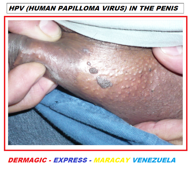
VPH, lesiones verrugosas en pene


PUBLICADO 1998, ACTUALIZADO 2017 - 2025
NOTA: PARA OBTENER TODA LA INFORMACIÓN DEBES LEER LOS ENLACES 


EDITORIAL ESPAÑOL 
===================
Hola amigos de la red DERMAGIC EXPRESS hoy te voy a poner una actualización del VIRUS DEL PAPILOMA HUMANO, VPH.
 
Hecho que inicie hace mas de 20 años con una publicación que salio a la red en el año 1999, con el nombre de EL VPH Y SU COMPORTAMIENTO EN NUESTRO MUNDO, donde hable del comportamiento de este virus en 43 países y estaba lejos la aparición de las VACUNAS contra el mismo.
 
En el año 2017 al 2023 hice una actualización y hoy 2025 te traigo dos versiones actualizadas del VIRUS DEL VPH, esta que estas leyendo y el enlace que te dejo mas abajo de este texto publicado con el nombre de ACTUALIZACIÓN DEL VIRUS DEL PAPILOMA HUMANO (2025).


1.) HISTORIA DEL VIRUS DEL VPH:
 
El primero en describir el virus del VPH fue el virologo y patologo Estadounidense Richard Shope en el año 1933, lo cual hizo en verrugas cutáneas de conejos. 

Posteriormente en el año de 1974,  el medico y virologo Aleman Harald zur Hausen fue el primero en proponer que el virus del VPH podría estar vinculado o relacionado con cancer cervical en humanos, lo cual fue cierto y le valió para ganar el Premio Nobel de Medicina en el año 2008

A partir de la década de los 80 y años siguientes, se investigo los distintos tipos de VPH, aislándolos y clasificándolos, siendo los primeros, los VPH1, 
VPH6, VPH11, VPH16 y VPH18,  este ultimo asociado a cancer cervical. 

2.) CARACTERÍSTICAS DEL VIRUS: 
 
 El virus del PAPILOMA HUMANO, VPH es un virus ADN de doble cadena, perteneciente a la familia Papillomaviridae.
 
Se caracteriza por ser un virus de tamaño pequeño y especifico de los humanos, el cual provoca multiples infecciones en la piel y mucosas, incluyendo verrugas y neoplasias intraepiteliales en el cuello uterino de las mujeres, muchas de las cuales evolucionan a cancer. 
 
3.) IMPORTANCIA: 
 
la Importancia de este virus esta dada por tres hechos:

A.- Es el virus de transmisión sexual más común en el mundo.

B.- Cerca del 80% de las personas sexualmente activas lo contraerán alguna vez.
 
C.-  Algunos Genotipos de VPH, como lo son el VPH16 y el VPH18 y otros mas, son altamente oncogénicos (cancerigenos), a los cuales se les atribuye la mayoría de los cánceres cervicouterinos.     
 
 
4.) CLASIFICACIÓN: (AÑO 2017-2023)
 
Para el año 2.023 hay aproximadamente 200 tipos de VPH, de los cuales 51 especies afectan la mucosa genital estando clasificados así:

A.) Mas carcinogénicos los tipos: 16, 18, 31, 33, 35, 39, 45, 51, 52, 56, 58, 59, 68, 73, y 82. 

B.) Probable alto riesgo: el 26, 53,y 66.

C.) Bajo riesgo cancerígeno: 6, 11, 40, 42, 43, 44, 54, 61, 70, 72, 73, 81, y 108.

5.) CLASIFICACIÓN ACTUAL (AÑO 2025):
 
 En el año 2012 la Asociación Internacional para la Investigación del cancer decide modificar esta clasificación, y hoy dia se consideran solo dos (2) tipos: los de ALTO RIESGO y los DE BAJO RIESGO, en este enlace encentras la actualización del VIRUS DEL VPH, PAPILOMA HUMANO, ACTUALIZACIÓN (2025) con mas información y Referencias bibliográficas.  

A.- ALTO RIESGO: El VPH16 y VPH18 siguen siendo los más comunes en cuanto a malignidad  representando entre el 70 - 75%  de los casos de cáncer cervicouterino a nivel mundial; otros sitios de localización son: ano, vulva, vagina, pene y orofaringe.

B.-Entre el 15 al 20% adicional de casos de malignidad están representados por los genotipos de ALTO RIESGO: VPH31, 33, 45, 52 y 58, que en conjunto contribuyen a un 15-20% adicional de los casos. 
 
C.- Los otros Genotipos  que destacan por ser del ALTO RIESGO, con un 5% de los casos son: VPH35, 59, 39, 56, 51, 68, 73, 26, 69 y 82, con algunas variaciones regionales, tal es el caso de áfrica donde prevalece el VPH35. 

D.- En cuanto a los Genotipos de BAJO RIESGO, los VPH6 y VPH11 son los mas frecuentemente reportados en la papilomatosis respiratoria recurrente y y en las verrugas anogenitales, NO ASOCIADOS A CANCER.
 
 Se decidió incluir a los subtipos de MODERADO RIESGO en el grupo de los de ALTO RIESGO, basados los científicos, en el hecho de que estos finalmente iban a evolucionar a MALIGNIDAD.
 
Todo esto que estas leyendo codujo a los laboratorios a buscar y crear VACUNAS para proteger a la población mundial de la proliferación de este VIRUS, adquirido 99,9% por transmisión sexual.
 
6.) HISTORIA de la creación de las VACUNAS: 
 
A.- En la década de 1980 comenzó la investigación para la creación de una vacuna, tras descubrirse la relación existente entre el virus del VPH con el cáncer cervical.
 
B.- En 1991, Ian Fraser y Jian Zhou (Universidad de Queensland, Australia) lograron desarrollar una tecnología para crear partículas similares al virus (VLPs), elemento fundamental para la posible creación de una VACUNA.
 
Estas partículas (VLPs) fueron creadas mediante ingeniería genética con la finalidad de estimular el sistema inmune, sin contenido de material genético viral infeccioso.

 C.- En 2006 la primera vacuna fue aprobada en Estados Unidos, por los laboratorios: 
Merck & Co, (EE.UU.), MSD en Europa y Sanofi-Pasteur, con el nombre comercial de GARDASIL (tetravalente), la cual protege contra los VPH6, VPH11, VPH16, y VPH18   para cáncer cervical,  y VPH6 y VPH11 para verrugas genitales.

D.- En el año 2007 fua aprobada por la FDA la VACUNA CERVARIX (bivalente) del laboratorio 
GlaxoSmithKline (GSK, Reino Unido), la cual protege contra los virus VPH16, y VPH18, para evitar fundamentalmente el cáncer cervical, con una cobertura de aproximadamente del 66%, y a la cual también se la atribuye "ALGO" de protección cruzada contra otros tipos de alto riesgo.
 
E.) En el año 2014-2015 fua aprobada la VACUNA GARDASIL-9, (nonavalente), una version mas amplia de la clásica  GARDASIL con protección contra los virus: VPH6, VPH11, VPH16, VPH18, VPH31, VPH33, VPH45, VPH52, VPH58, es decir con una mayor cobertura (90%), contra los tipos de VPH de ALTO RIESGO de cáncer cervical y verrugas.
 
 
 Los efectos adversos, los cuales suelen ser transitorios y autolimitados, más frecuentes son:
 
A.- Locales y leves:
 
- Dolor.
- Eritema (enrojecimiento) e hinchazón en el sitio de la inyección. 
- Cefalea.
- Mareos. 
 
B .- Moderados a graves: 
 
- Sincope y desmayos: especialmente en adolescentes, por lo que se recomienda observar al  paciente, por lo menos 15 minutos luego de la administración, con el objetivo de prevenir lesiones secundarias, en caso de desmayos.
 
- Pueden ocurrir reacciones anafilacticas graves cuando hay alergia comprobada a los componentes de la vacuna, y son extremadamente raras. 
 
 - La incidencia de Sincope y reacciones locales se ha descrito con mayor frecuencia en la GARDASIL-9 (nonavalente), en comparación con la clásica GARDASIL (tetravalente).
 
-  Hay ABSOLUTA contraindicacion , cuando se ha comprobado que el paciente ha experimentado reacciones severas, a la administración de otras vacunas. 
 
C.- Eventos adversos posteriores a la vacuna contra el virus del papiloma humano 9-valente (GARDASIL® 9) notificados al Sistema de Notificación de Eventos Adversos de Vacunas (VAERS), 2015-2024:
 
Eventos no mencionados en la ETIQUETA del producto:
 
 
Hubo ademas 57 informes de defunción (MUERTE), de los cuales 18 fueron en mujeres embarazadas, pero los términos o características neurológicas carecían de patrones etológicos contundentes, de modo que el sistema VAERS, concluye que dicha vacuna es "segura".
 
Pero si quieres indagar mas a fondo de lo que ha sucedido con estas vacunas aquí te dejo el enlace de la publicación EL SÍNDROME POST VACUNACIÓN GARDASIL Y CERVARIX (2017), el cual "muestra" una cara totalmente a el sistema VAERS del año 2015 al 2024.

 8.) CONCLUSIÓN:
"En un periodo de 23 a 25 años aparecieron unas 120 CEPAS nuevas DEL VIRUS PAPILOMA HUMANO V.P.H, quizá este aumento cabalgante fue lo que llevo a los laboratorios a FABRICAR VACUNAS para esta enfermedad, la experiencia con las mismas ha sido CONTROVERSIAL", pero el hecho de que protejan contra los dos tipos de VPH mas oncogenicos es un "SIGNO POSITIVO."

En este enlace encuentras la publicación ORIGINAL del VIRUS DEL VPH Y SU COMPORTAMIENTO EN EL MUNDO (1999); es importante que te la leas, para que aprendas como ha evolucionado el comportamiento de este virus en la población mundial desde ese año hasta nuestros días. 

NOTA: Actualmente estas vacunas tienen un costo de 250$ dolares Americanos en Venezuela. 

Saludos a todos!!!

Dr. José Lapenta R.
Dr. José M. Lapenta R.,


EDITORIAL ENGLISH
=================
Hello friends of the DERMAGIC EXPRESS network, today I'm going to give you an UPDATE ON THE HUMAN PAPILLOMAVIRUS (HPV).

This article began more than 20 years ago with a publication published online in 1999, entitled "HPV AND ITS BEHAVIOR IN OUR WORLD." I discussed the behavior of this virus in 43 countries, and the emergence of vaccines against it was still far from complete.

From 2017 to 2023, I updated it, and today, in 2025, I'm bringing you two updated versions of the HPV virus: the one you're reading and the link at the end of this article, published under the name HUMAN PAPILLOMA VIRUS (HPV) UPDATE (2025)."

1.) HISTORY OF THE HPV VIRUS:


The first to describe the HPV virus was the American virologist and pathologist Richard Shope in 1933, which he did in rabbit skin warts.

Later, in 1974, the German physician and virologist Harald zur Hausen was the first to propose that the HPV virus could be linked or related to cervical cancer in humans, which proved true and earned him the Nobel Prize in Medicine in 2008.

Beginning in the 1980s and following years, the different types of HPV were investigated, isolated, and classified. The first were HPV1, HPV6, HPV11, HPV16, and HPV18, the latter being associated with cervical cancer.

2.) CHARACTERISTICS OF THE VIRUS:


The human papillomavirus (HPV) is a double-stranded DNA virus belonging to the Papillomaviridae family.

It is characterized by being a small, human-specific virus that causes multiple skin and mucous membrane infections, including warts and cervical intraepithelial neoplasia in women, many of which progress to cancer.

3.) IMPORTANCE:
The importance of this virus is based on three facts:

A. It is the most common sexually transmitted virus in the world.

B. About 80% of sexually active people will contract it at some point.

C.- Some HPV genotypes, such as HPV16 and HPV18, among others, are highly oncogenic (carcinogenic), and are responsible for the majority of cervical cancers.

4.) CLASSIFICATION: (YEAR 2017-2023):

As of 2023, there are approximately 200 types of HPV, of which 51 species affect the genital mucosa. They are classified as follows:

A.)Mostcarcinogenic types
 HPV16, HPV18, HPV31, HPV33, HPV35, HPV39, HPV45, HPV51, HPV52, 
 HPV56, HPV58, HPV59, HPV68, HPV73, and HPV82.

B.) Probable high risk: 
HPV26, HPV53, and HPV66.

C.) Low carcinogenic risk: 
HPV6, HPV11, HPV40, HPV42, HPV43,HPV HPV44,
 HPV54, HPV61, HPV70, HPV72, HPV73, HPV81, and HPV108.

5.) CURRENT CLASSIFICATION (YEAR 2025):

In 2012, the International Association for Cancer Research decided to modify this classification, and today only two (2) types are considered: HIGH RISK and LOW RISK. At this link, you will find the update on the HPV, HUMAN PAPILLOMA VIRUS, UPDATE (2025). 
with more information and bibliographic references.

A.- HIGH RISK: HPV16 and HPV18 continue to be the most common types of malignancy, representing between 70-75% of cervical cancer cases worldwide. Other sites include: anus, vulva, vagina, penis, and oropharynx.

B. Between 15 and 20% of additional malignancy cases are represented by the HIGH-RISK genotypes: HPV31, 
HPV33, HPV45, HPV52, and HPV58, which together contribute to an additional 15-20% of cases.

C. The other genotypes that stand out as HIGH-RISK, accounting for 5% of cases, are: HPV35, 
HPV59, HPV39, HPV56, HPV51, HPV68, HPV73, HPV26, 
HPV69, and HPV82, with some regional variations, such as in Africa, where HPV35 is prevalent.

D.- Regarding the LOW-RISK genotypes, HPV6 and HPV11 are the most frequently reported in recurrent respiratory papillomatosis and anogenital warts, NOT ASSOCIATED WITH CANCER.

It was decided to include the MODERATE RISK subtypes in the HIGH RISK group, based on the scientists' assumption that these would eventually evolve into MALIGNANCY.

All of this you are reading led laboratories to search for and create VACCINES to protect the world's population from the spread of this VIRUS, 99.9% of which is acquired through sexual transmission.

6.) HISTORY OF THE CREATION OF VACCINES:

A.- In the 1980s, research into the creation of a vaccine began after the link between the HPV virus and cervical cancer was discovered.

B.- In 1991, Ian Fraser and Jian Zhou (University of Queensland, Australia) developed a technology to create virus-like particles (VLPs), a fundamental element for the possible creation of a VACCINE.

These particles (VLPs) were created through genetic engineering with the aim of stimulating the immune system, without containing infectious viral genetic material.

C.- In 2006, the first vaccine was approved in the United States by the laboratories: Merck & Co. (USA), MSD in Europe, and Sanofi-Pasteur, under the brand name GARDASIL (tetravalent). It protects against HPV6, HPV11, HPV16, and HPV18 for cervical cancer, and HPV6 and HPV11 for genital warts.

D.- In 2007, the FDA approved the CERVARIX (bivalent) vaccine from GlaxoSmithKline (GSK, United Kingdom). It protects against the HPV16 and HPV18 viruses, primarily to prevent cervical cancer, with a coverage of approximately 66%. It is also attributed with "SOME" cross-protection against other high-risk types.

E.) In 2014-2015, the GARDASIL-9 VACCINE (nine-valent) was approved. This is a broader version of the classic GARDASIL vaccine, offering protection against the following viruses: HPV6, HPV11, HPV16, HPV18, HPV31, HPV33, HPV45, HPV52, and HPV58. This vaccine offers greater coverage (90%) against high-risk HPV types for cervical cancer and warts.

7.) SIDE EFFECTS OF THE VACCINES:

The most common adverse effects, which are usually transient and self-limiting, are:

A.- Local and mild:

- Pain.
- Erythema (redness) and swelling at the injection site.
- Headache.
- Dizziness.

B. Moderate to severe:


- Syncope and fainting: especially in adolescents, so it is recommended that the patient be observed for at least 15 minutes after administration to prevent secondary injuries in the event of fainting.

- Severe anaphylactic reactions can occur when there is a known allergy to vaccine components and are extremely rare.

- The incidence of syncope and local reactions has been reported more frequently with GARDASIL-9 (nine-valent) compared to the classic GARDASIL (four-valent).

- There is an ABSOLUTE contraindication to the administration of other vaccines when the patient has been known to experience severe reactions.

C. Adverse events following the 9-valent human papillomavirus vaccine (GARDASIL® 9) reported to the Vaccine Adverse Event Reporting System (VAERS), 2015-2024:

Events not listed on the product label:

- Postural orthostatic tachycardia syndrome.
- Eye movement disorders.
- Autoimmune thyroiditis.
- Postural abnormality.

There were also 57 reports of deaths, 18 of which were in pregnant women, but the neurological characteristics or features lacked compelling etiological patterns, so the VAERS system concludes that the vaccine is "safe."

But if you want to delve deeper into what has happened with these vaccines, here is the link to the publication "POST-VACCINATION SYNDROME WITH GARDASIL AND CERVARIX" (2017), which completely "shows" the other "face" of the VAERS system from 2015 to 2024.

8.) CONCLUSION:
"Over a period of 23 to 25 years, some 120 new STRAINS OF THE HUMAN PAPILLOMAVIRUS (HPV) appeared. Perhaps this dramatic increase was what led laboratories to manufacture vaccines for this disease. Experience with these vaccines has been CONTROVERSIAL," but the fact that they protect against the two most oncogenic HPV types is a "POSITIVE SIGN."
At this link, you'll find the original publication "HPV VIRUS AND ITS BEHAVIOR IN THE WORLD" (1999). It's important to read it to learn how the behavior of this virus has evolved in the global population from that year to the present.

NOTE: These vaccines currently cost $250 in Venezuela.

Greetings to all!!!

Dr. José Lapenta R.
Dr. José M. Lapenta R.,


=======================================================================
REFERENCIAS BIBLIOGRÁFICAS/ BIBLIOGRAPHICAL REFERENCES
=======================================================================
 A.- Adverse events following 9-valent human papillomavirus vaccine (GARDASIL 9) reported to the Vaccine Adverse Event Reporting System (VAERS), 2015-2024.
=======================================================================
1.) Detection and typing of human papillomaviruses by polymerase chain reaction in cervical scrapes of Croatian women with abnormal cytology. (CROATIA)
2.) Risk factors for HPV DNA detection in middle-aged women. (FRANCE)
3.) Human papillomavirus 16 and 18 infection of the uterine cervix in women with different grades of cervical intraepithelial neoplasia (CIN). (SLOVENIA)
4.) HPV 16 infection and progression of cervical intra-epithelial
neoplasia: analysis of HLA polymorphism and HPV 16 E6 sequence variants. (AMSTERDAM)
5.) HPV prevalence among Mexican women with neoplastic and normal cervixes. (MEXICO)
6.) Cigarette smoking and high-risk HPV DNA as predisposing factors for high-grade cervical intraepithelial neoplasia (CIN) in young Brazilian women. (BRAZIL)
7.) Prevalence of human papillomavirus infection in premalignant and malignant lesions of the oral cavity in U.K. subjects: a novel method of detection. (LONDON)
8.) Causes of cervical cancer in the Philippines: a case-control study. (PHILLIPINES)
9.) Novel HPV types present in oral papillomatous lesions from patients with HIV infection. (GERMANY)
10.) HPV-types, cytological and histopathological findings in three groups of women with possible HPV-related disease. (SWEDEN)
11.) A general primer GP5+/GP6(+)-mediated PCR-enzyme immunoassay method for rapid detection of 14 high-risk and 6 low-risk human papillomavirus genotypes in cervical scrapings. (AMSTERDAM)
12.) Prevalence of antibodies to human papillomavirus (HPV) type 16 virus-like particles in relation to cervical HPV infection among college women. (USA)
13.) Detection of human papillomavirus mRNA and cervical cancer cells in peripheral blood of cervical cancer patients with metastasis. (TAIWAN)
14.) Human papillomavirus in tissue of bladder and bladder carcinoma specimens. A preliminary study. (GERMANY)
15.) Detection of human papillomavirus (HPV) type 47 DNA in malignant lesions from epidermodysplasia verruciformis by protocols for precise typing of related HPV DNAs. (JAPAN)
16.) HLA and susceptibility to cervical neoplasia. (NETHERLANDS)
17.) Non-isotopic in situ hybridization of HPV types in cervical intraepithelial lesions in patients with AIDS. (BRAZIL)
18.) HLA-A2-restricted peripheral blood cytolytic T lymphocyte response to HPV type 16 proteins E6 and E7 from patients with neoplastic cervical lesions. (GERMANY)
19.) Human papilloma virus 16-18 infection and cervical cancer in Mexico: a case-control study. (MEXICO)
20.) Detection of human papillomavirus (HPV) type 6, 16 and 18 in head and neck squamous cell carcinomas by in situ hybridization. (CROATIA)
21.) Prevalence of human papillomavirus infection in women attending a sexually transmitted disease clinic. (JAPAN)
22.) Demonstration of multiple HPV types in laryngeal premalignant lesions using polymerase chain reaction and immunohistochemistry. (ITALY)
23.) Adenocarcinoma of the uterine cervix in Ireland and Sweden: human papillomavirus infection and biologic alterations. (IRELAND AND SWEDEN)
24.) Risk factors for high-risk type human papillomavirus infection among Mexican-American women. (USA-MEXICO)
25.) Many different papillomaviruses have low transcriptional activity in spite of strong epithelial specific enhancers. (SINGAPORE)
26.) Low frequency of human papillomavirus infection in initial papillary bladder tumors. (CANADA)
27.) Human papillomavirus genotype spectrum in Czech women: correlation of HPV DNA presence with antibodies against HPV-16, 18, and 33 virus-like particles. (CZECH REPUBLIC)
28.) Mucosal oncogenic human papillomaviruses and extragenital Bowen disease. (FRANCE) 29.) High prevalence of a variety of epidermodysplasia verruciformis-associated human papillomaviruses in psoriatic skin of patients treated or not treated with PUVA. (GERMANY)
30.) Human papillomavirus type 31 oncoproteins E6 and E7 are required for the maintenance of episomes during the viral life cycle in normal human keratinocytes. (USA)
31.) Clinical, histopathologic, and molecular aspects of cutaneous human papillomavirus infections. (USA)
32.) Screening for genital human papillomavirus: results from an international validation study on human papillomavirus sampling techniques. (SPAIN)
33.) Use of the same archival papanicolanou smears for detection of human papillomavirus by cytology and polymerase chain reaction. (AUSTRALIA)
34.) Detection and typing of human papillomavirus in cervical cancer in the Thai. (THAILAND)
35.) Correlation between polymerase chain reaction and cervical cytology for detection of human papillomavirus infection in women with and without dysplasia. (NORWAY)
36.) Detection and quantitation of human papillomavirus by using the fluorescent 5' exonuclease assay. (SWEDEN)
37.) Risk factors for HPV detection in archival Pap smears. A population-based study from Greenland and Denmark. (GREENLAND AND DENMARK)
38.) [Human papillomavirus infection in women with and without abnormal cervical cytology]. (MEXICO)
39.) Follow-up of human papillomavirus (HPV) DNA and local anti-HPVantibodies in cytologically normal pregnant women. (HUNGARY)
40.) Relatively low prevalence of human papillomavirus 16, 18 and 33 DNA inthe normal cervices of Japanese women shown by polymerase chain reaction. (JAPAN)
41.) Comparison of a one-step and a two-step polymerase chain reaction with degenerate general primers in a population-based study of human papillomavirus infection in young Swedish women. (SWEDEN)
42.) Human papillomavirus DNA in unselected pregnant and non-pregnant women. (FINLAND)
43.) Prevalence of HPV cervical infection in a family planning clinic determined by polymerase chain reaction and dot blot hybridisation. (LONDON)
44.) Type-specific prevalence of human papillomavirus DNA among Jamaican colposcopy patients. (JAMAICA)
45.) Polymerase chain reaction detection and restriction enzyme typing of human papillomavirus in cervical carcinoma. (MALAYSIA)
46.) Human papillomavirus (HPV) cervical lesions: results from 300 Italian women studied with DNA hybridization techniques and morphology. (ITALY)
47.) Detection of human papillomavirus (HPV) DNA in human prostatic tissues by polymerase chain reaction (PCR). (USA)
48.) High-risk human papillomavirus types in cytologically normal cervical scrapes from Kenya. (KENYA)
49.) Detection of type specific human papillomavirus (HPV) DNA in cervical cancers of Indian women. (INDIA)
50.) Natural history of cervical human papillomavirus lesions. (JAPAN)
51.) Detection of human papillomavirus types in cervical lesions of patients from Taiwan by the polymerase chain reaction. (TAIWAN)
52.) Human papillomaviruses in cervical cancer I. HPV-16 and 18 predominate in the Greek population. (TAIWAN-CHINA) 53.) The prevalence of cervical infection with human papillomaviruses and cervical dysplasia in Alaska Native women. (ALASKA)
54.) Detection of human papillomaviruses in exfoliated cervicovaginal cells by in situ DNA hybridization analysis. (TAIWAN)
55.) Detection and typing of human papillomavirus in cervical specimens of Turkish women. (TURKEY)
56.) Prevalence of human papillomavirus DNA in cervical tissue. Retrospective analysis of 855 cervical biopsies. (GERMANY)
57.) Prevalence of human papillomavirus DNA in female cervical lesions from Rio de Janeiro, Brazil. (BRAZIL)
58.) Prevalence of human papilloma virus 16 or 18 in cervical cancer in Hualien, eastern Taiwan. (TAIWAN)
59.) Human papillomavirus types 52 and 58 are prevalent in cervical cancers from Chinese women. (CHINA)
60.) Human papillomavirus infection and risk determinants for squamous intraepithelial lesion and cervical cancer in Japan. (JAPAN)
61.) Detection and typing of human papillomavirus in cervical carcinomas in Russian women: a prognostic study. (RUSSIA)
62.) Serologic response to human papillomavirus type 16 (HPV-16) virus-like particles in HPV-16 DNA-positive invasive cervical cancer and cervical intraepithelial neoplasia grade III patients and controls from Colombia and Spain. (COLOMBIA, SPAIN)
63.) Detection of human papillomavirus-related oral verruca vulgaris among Venezuelans. (VENEZUELA)
64.) Oncogenic association of specific human papillomavirus types with cervical neoplasia. (USA, PERU and BRAZIL)
65.) Chromosome fragility in lymphocytes of women with cervical uterine lesions produced by human papillomavirus. (ECUADOR)
66.) Multifocal papilloma virus epithelial hyperplasia [see comments] (GUATEMALA)
67.) Genital human papillomavirus infection in Panama City prostitutes. (PANAMA)
68.) Risk factors for genital papillomavirus infection in populations at high and low risk for cervical cancer. (PANAMA)
69.) Demonstration of multiple HPV types in normal cervix and in cervical squamous cell carcinoma using the polymerase chain reaction on paraffin wax embedded material. (ENGLAND)
70.) Morphological correlation of human papillomavirus infection of matched cervical smears and biopsies from patients with persistent mild cervical cytological abnormalities. (ENGLAND)
71.) Epidermodysplasia verruciformis in Africans. (SOUTH AFRICA)
72.) Transmissibility and treatment failures of different types of human papillomavirus. (ISRAEL)
73.) High frequency of detection of epidermodysplasia verruciformis-associated human papillomavirus DNA in biopsies from malignant and premalignant skin lesions from renal transplant recipients. (THE NETHERLANDS)
===================================================================

Si Te ha gustado, Compartelo